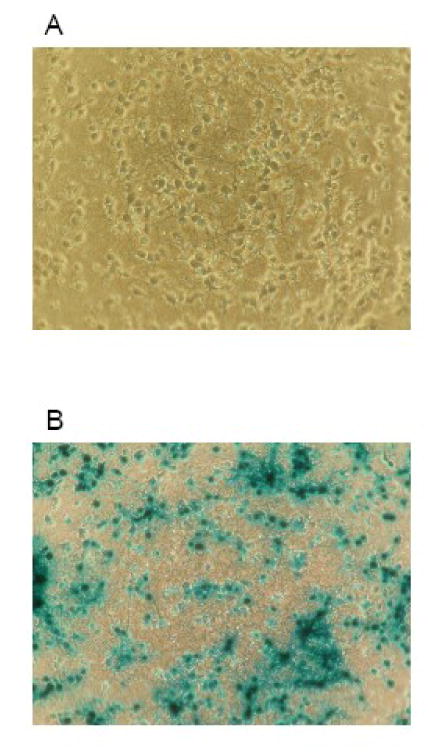
Figure 1. Photomicrographs of cultured cortical neurons before and after infection with the adenoviral vector Ad5CMVntLacZ.

Panel A shows cortical neurons in culture on day 9 in vitro. On panel B, cells were incubated with the Ad5CMVntLacZ vector in the regular culture medium for 24 h using a titer of 10 plaque forming units per cell. Thereafter, the cells were fixed and stained for β-galactosidase using a commercially available kit. Cells were counted in 5 randomly selected fields in each sample using a phase contrast microscope to calculate the ratio of cells expressing the desired protein.
