
Figure 2. Adenoviral catalase gene transfer enhanced neuronal catalase activity and expression but did not influence the expression of other antioxidants.
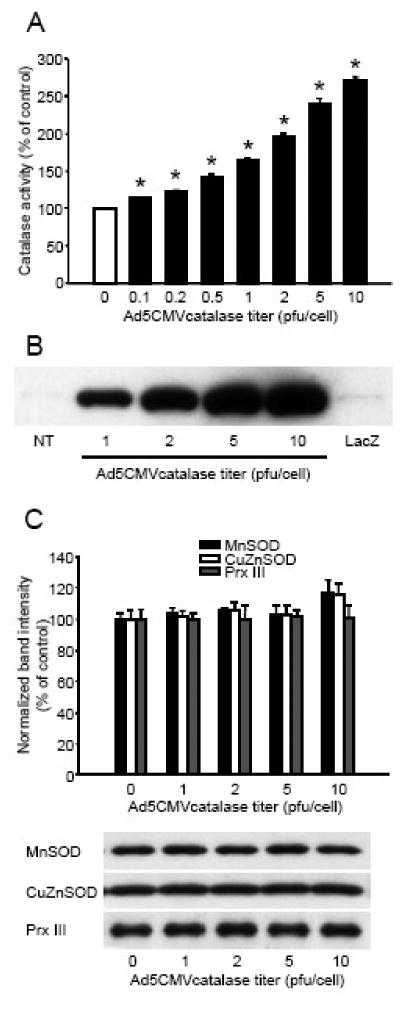
Cultured cortical neurons were incubated with different titers of the Ad5CMVcatalase vector or 10 plaque forming units/cell titer of the Ad5CMVntLacZ (LacZ) vector for 24 h in the regular cell culture medium. The cultures were rinsed and catalase enzyme activity was measured using a commercially available kit (Panel A). Enzyme activity was expressed as percent of non-infected control. *Significant difference (p<0.05) compared with untreated control. Data are expressed as mean ± SEM; n = 16 in each group. In other experiments, 24 h after infection, the cultures were rinsed and protein was extracted and subjected to western blot analysis for catalase (Panel B), or manganese dependent superoxide dismutase (MnSOD), copper and zinc dependent superoxide dismutase (CuZnSOD), and peroxiredoxin III (Prx III) (Panel C). Bands were normalized to the intensity of β-actin, and were expressed as percent of non-infected control. Representative blots are shown below the graph. Data are expressed as mean ± SEM; n = 4 in each group; NT, non-treated; pfu, plaque forming unit.