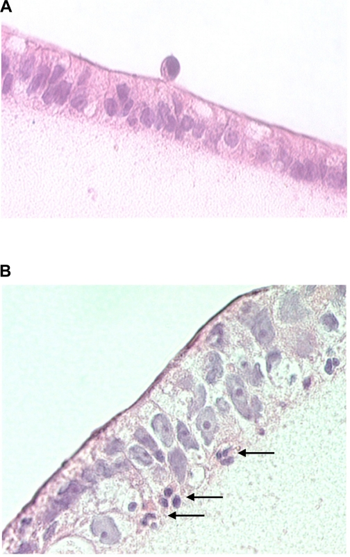
Fig. 1.
Histological sections of T84 cell monolayers incubated with neutrophils [polymorphonuclear neutrophils (PMN)]. In A, PMN were placed on the apical side of T84 monolayers. B: appearance 30 min after adding N-formyl-methionyl-leucyl-phenylalanine (fMLP) to the basolateral side. Marked epithelial transmigration of PMN is apparent (arrows).

