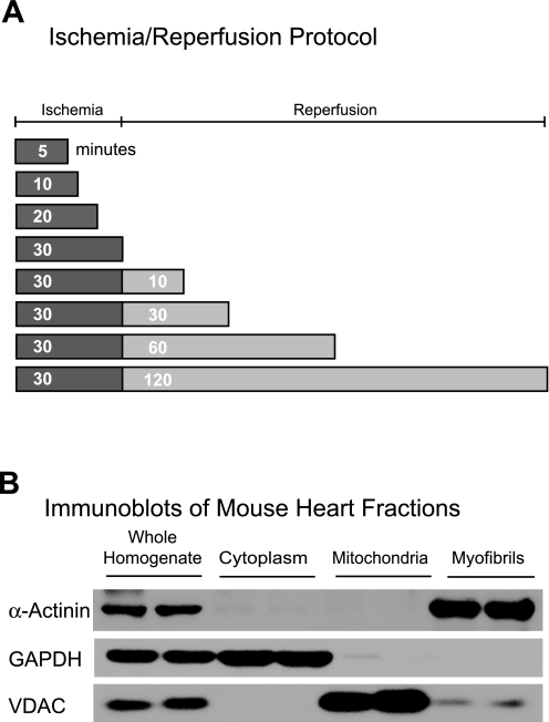
Fig. 2.
Diagram of the ischemia-reperfusion (I/R) protocol used in this study and subcellular fractionation. A: after 30 min of equilibration mouse hearts were subjected to various times of I/R on a Langendorff apparatus (n = 3 mouse hearts/time). A continual perfusion control that matched each ischemia and I/R time was also generated. B: after each treatment, hearts were homogenized and, after removal of a sample for later analysis, homogenates were subjected to subcellular fractionation. Tissue was later homogenized and fractionated by differential centrifugation and examined by immunoblot for α-actinin, GAPDH, and voltage-dependent anion channel (VDAC), which are marker proteins for myofibrils, cytosol, and mitochondria, respectively.

