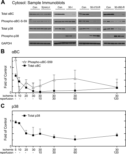
Fig. 4.
Effect of ischemia and I/R on αBC in cytosolic fractions. Immunoblotting was performed on cytosolic fractions from all the time points to examine total αBC and GAPDH. After subcellular fractionation, samples of the cytosolic fractions from the 8 experimental samples shown in Fig. 1A, and 8 time-matched controls (n = 3 hearts per time), were subjected to immunoblot analysis. A: samples from 4 of the time points were analyzed by immunoblotting for total αBC, phospho-αBC-S59, p38, phospho-p38, and GAPDH, as labeled. B: immunoblots of all samples for total αBC (▪) and phospho-αBC-S59 (▵) were quantified; values are shown as fold over control hearts for each time point normalized to VDAC. C: immunoblots of all samples for total p38 (▪) were quantified; values are shown as fold over control hearts for each time point normalized to VDAC. All values are means ± SE; n = 3 for each time point. All values are significantly greater than control perfusion values (P < 0.05). In cases where error bars are not visible, they are smaller than the symbol. Due to the lack of signal in control lanes, it was not possible to determine the proportion of total p38 that was phosphorylated under control conditions, so only total p38 is shown in this figure.

