Fig. 3.
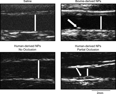
Representative images of carotid arteries obtained by B-mode ultrasonography. Patent, injured arteries of rabbits inoculated with saline (top, left), bovine-derived NPs (top, right), and human-derived NPs (bottom, left) are shown at 5 wk postinjury. An injured artery of a rabbit inoculated with human-derived NPs is shown at 2 wk postsurgery (bottom, right); this artery was occluded by week 3. Bars designate the lumens. A stitch closing access to the artery following endothelial denudation was used as a marker of the injured area (arrow; top, right). Intimal thickening in the artery that subsequently occluded (arrow; bottom, right) is shown. Scale bar is 2 mm.

