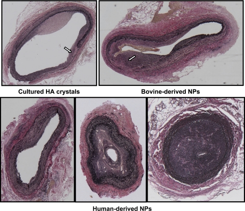
Fig. 4.
Light micrographs of representative sections of injured carotid arteries from rabbits injected with HA crystals exposed to culture medium (top, left), bovine-derived NPs (top, right), and human-derived NPs (bottom). Sections are stained with an elastin van Giesen stain and shown at ×5 magnification. All arteries were collected at 35 days postoperatively. Uninjured arteries from all rabbits are indistinguishable from the uninjured artery shown in Fig. 2. Arrows indicate areas of disrupted internal elastic lamina/media layer.

