Fig. 4.
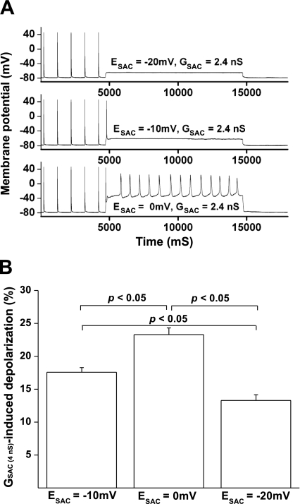
The effect of SAC reversal potential (ESAC) on GSAC-induced cell depolarization and automaticity in rat ventricular myocytes. A: at a relatively low value of GSAC (2.4 nS), increasing ESAC levels from −20 to 0 mV significantly increased the GSAC-induced cell depolarization. The sustained automaticity was successfully induced by the GSAC at ESAC = 0 mV, but not at the ESAC value of −10 or −20 mV. B: bar graph showing the cell depolarization induced by 2.4-nS GSAC at the ESAC level of −20, −10, and 0 mV (n = 6).

