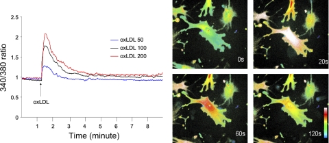
Fig. 4.
Oxidized low-density lipoprotein (oxLDL)-induced Ca2+ release in endothelial cells from B6 mice. Left: changes over time in cytosolic Ca2+ levels after stimulation with various doses of oxLDL. The increase in the ratio of fluorescence at wave length 340 nm to 380 nm indicates a rise in intracellular free Ca2+. oxLDL induced a dose-dependent increase in Ca2+ levels. Right: confocal microscopy images showing changes in intracellular distribution of Ca2+ labeled with fluorescent dye fura 2 at 0, 20, 60, and 120 s in endothelial cells from B6 mice after addition of oxLDL.

