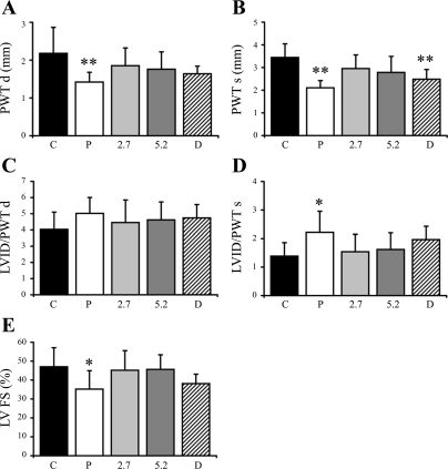
Fig. 2.
Echocardiographic data. PWTd, posterior wall thickness in diastole (A); PWTs, posterior wall thickness in systole (B); LVID/PWTd, ratio of left ventricle (LV) internal dimension to posterior wall thickness in diastole (C); LVID/PWTs, ratio of LV internal dimension to posterior wall thickness in systole (D). FS, fractional shortening (E). Values are means ± SD. **P < 0.01 vs. control; *P < 0.05 vs. control.

