Fig. 2.
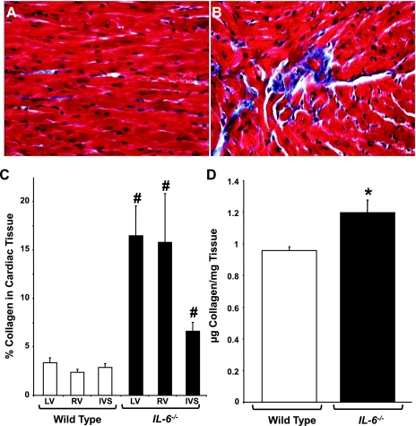
Ventricular collagen levels in IL-6−/− mice. A and B: representative Masson's trichrome-stained sections from WT and IL-6−/− LV chambers, respectively. C: quantification of collagen content in the LV and RV free walls and intraventricular septa (IVS). D: quantification of cardiac collagen content via hydroxyproline analysis in adult mice. N = 4–6 animals per condition. #P < 0.001 and *P < 0.05 vs. WT.

