Figure 1.
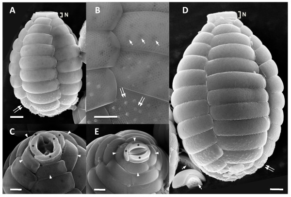
SEM images of photosynthetic Paulinella strains FK01 (A – C) and M0880/a (D, E). FK01 is smaller in cell size than M0880/a (i.e., the scale bar is the same in A and D). Distinctive, multiple fine pores are present on the surface of scales of FK01 (B). The five (C) oral scales (asterisks) are shown from FK01, whereas only three (E) are found in M0880/a. The projecting oral scales (N), first row of body scales (arrow head), "sieve-plate" in internal surface (arrow), and pustules (double arrow) are indicated. Scale bars (A, D, E = 2 μm; B, C = 1 μm).

