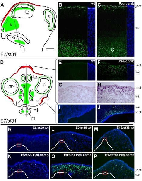
Figure 3. SOX5 immunostaining in Pea-comb and wild-type embryonic heads.
(A, D) Schematic drawings of sagittal and cross-sections of an E7 chick head. Green indicates SOX5 immunostainings that are identical in wild-type and Pea-comb birds, red indicates SOX5 staining unique for Pea-comb. The planes of the drawings are shown as shaded lines. Scale bar 1 mm. (B, E, I) Fluorescence micrographs of the wattle and comb regions with SOX5 immuno- and DAPI nuclear staining of E7 wild-type and (C, F, J) Pea-comb birds. (G, H) Bright-field micrographs of cRNA in situ hybridization for SOX5 mRNA in wild-type and Pea-comb. The positions of the comb and wattle regions shown in panels B, C, E–J are boxed in the schematic drawings. Scale bars 100 μm. (K–P) SOX5 immuno- and DAPI nuclear staining in the comb region of E6, E9 and E12 wild-type and Pea-comb chickens. Insets show schematic drawings of the comb-ridge shapes in wild-type and Pea-comb. The positions of corresponding fluorescence micrographs are boxed. Scale bar 100 μm. ect; ectoderm, e; eye, l; lumen of nostril, m; Meckel's cartilage, me; mesenchyme, nr; neural retina, o; optic lobe, s; interorbital septum, st; stage according to Hamburger and Hamilton [51], t; tongue, te; telencephalon, wt; wild-type.

