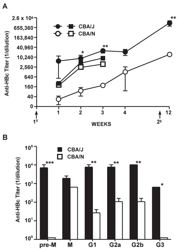
Figure 5.
Anti-HBc antibody production in vivo is influenced by B1a cells. (A). Groups of 5 wild-type CBA/J or CBA/N (xid) mice were immunized (1°) with HBcAg149 (20 μg in saline) and boosted (2°) (i.p.) with 10 μg of HBcAg149 in saline and serum anti-HBc antibody levels determined by ELISA at the indicated times (○-○, ●-●). Serum anti-HBc IgG antibody was quantitated by endpoint dilution of sera (titer). Groups of 3 mice each were also immunized (1°) with HBsAg (20 μg, IFA) and anti-HBs antibody determined by ELISA (□-□, ■- ■). (B). Groups of 3 CBA/J or CBA/N (xid) mice were immunized (i.p.) with HBcAg149 (20 μg insaline) and 4 weeks later serum IgM and IgG isotype- specific anti-HBc antibodies were determined by ELISA. Pre-M, pre-existing IgM anti-HBc (prior to immunization). Results represent the mean values (± SD) of individual mouse sera (*P = 0.02, **P = 0.005, ***P = 0.002).

