Fig. 11.
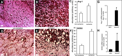
Immunohistochemical validation of 2 ischemia-sensitive candidate genes Arg-1 and mitochondria superoxide dismutase (SOD2). Wound-edge tissues were collected on day 3 (SOD2) or day 7 (Arg-1) after wounding. Formalin-fixed paraffin-embedded biopsy tissues were sectioned (5 μm) and immunostained [diaminobenzidine (DAB) detection with hematoxylin counterstain] with antibodies against either arginase 1 (anti-Arg-1) or SOD2 (anti-SOD2) after heat-induced epitope retrieval. The brown coloration indicates positive staining (A and B, D and E). C, F: the area of DAB stain was estimated using Adobe Photoshop 6.0 employing a color subtractive process. Data are means ± SD (n = 3). *P < 0.05. Expression of Arg-1 (G) and SOD2 (H) in human skin and wound-edge tissue collected from chronic wounds. Quantitative real-time PCR was performed. Data are mean ± SD (n = 4). *P < 0.05.

