Fig. 5.
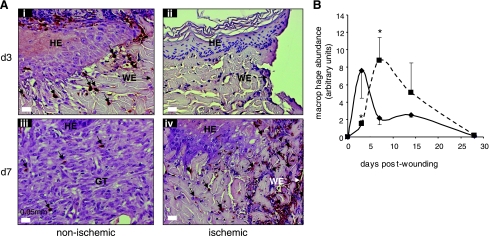
Dysregulated macrophage infiltration in ischemic wounds. Wound-edge tissues were collected at the indicated time points after wounding. Formalin-fixed paraffin-embedded biopsy tissues were sectioned (5 μm) and stained using L1 macrophage/calprotectin immunostaining (brown). A: representative images of ischemic (right) and nonischemic (left) wounds showing macrophages (brown). The tissue sections were counterstained with hematoxylin (blue). HE, hyperproliferative epithelium; GT, granulation tissue; WE, wound-edge orientation; B: kinetics of macrophage infiltration in the nonischemic (solid line, ⧫) and ischemic wounds (dotted line, ▪). Relative quantification (arbitrary units) of macrophages in the tissue sections obtained 3–28 days postwounding was performed using an image processing tool kit. Data are means ± SD (n = 3). *P < 0.05.

