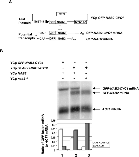
FIGURE 1.
Nab2 protein inhibits 3′-end formation of its own mRNA. (A) Diagram of the test plasmid, which allows transcription from the MET17 promoter of the inserted test gene fused to green fluorescent protein (GFP). CYC1pA designates the polyadenylation site of CYC1. The potential transcripts, distinguished by their different 3′-ends, are shown below the diagram of the test plasmid. (B) Strain YSB501 (YCpNAB2) or YSB502 (YCpnab2-1) carrying the indicated plasmids was grown in SCD–URA-LEU at 30°C; total RNA was isolated and the levels of GFP-NAB2 transcripts determined by Northern blot analysis with an oligonucleotide probe complementary to the GFP portion of the mRNA. YCpSL-GFP-NAB2-CYC1 designates the use of the reporter plasmid with the stem–loop insertion in the 5′-UTR of the reporter mRNA. The bar graph below reports the ratio of the GFP-fusion transcripts to ACT1 mRNA and is based on two independent measurements.

