Figure 3.
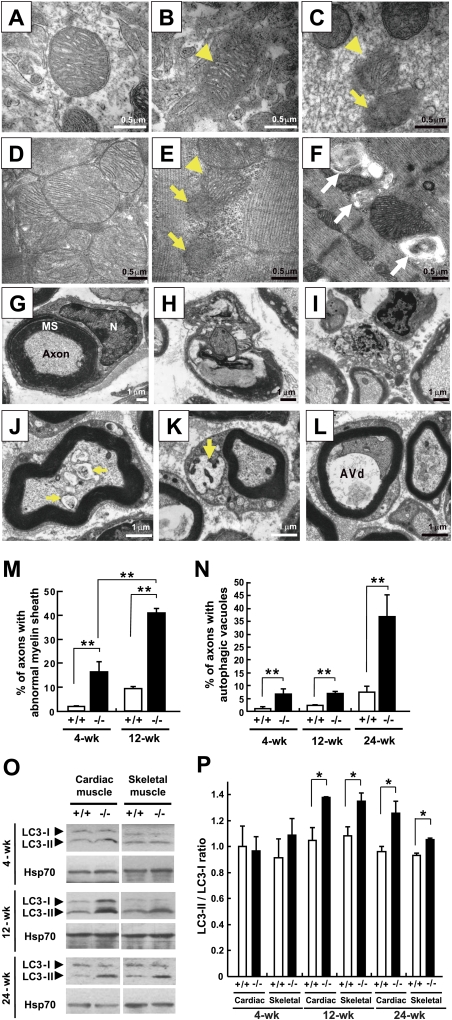
Mitochondrial degeneration and autophagy induction in the muscles and neurons of the Cisd2−/− mice. (A) Wild-type mitochondria in the brain (hippocampus). (B) A Cisd2−/− mitochondrion in the brain (hippocampus). Note that the outer mitochondrial membrane has broken down (arrowhead), while the inner cristae appear to be intact. (C) Cisd2−/− mitochondria in sciatic nerve. One mitochondrion (arrowhead) has a destroyed OM, but with cristae still visible; the other mitochondrion (arrow) has destroyed OMs and IMs. (D) Wild-type mitochondria in cardiac muscle. (E) Cisd2−/− mitochondria in cardiac muscle. This micrograph shows one mitochondrion (arrowhead) with a destroyed OM and two degenerated mitochondria consisting of debris (arrows). (F) A cluster of autophagic vacuoles and abnormal mitochondria was observed between the myofibrils of Cisd2−/− skeletal muscle (white arrows). (G) A wild-type myelinated axon of the sciatic nerve. (N) Nucleus of Schwann cell; (MS) myelin sheath. (H) A myelinated axon of sciatic nerve from a Cisd2−/− mouse. An ovoid with a disintegrating myelin sheath and a degenerating axonal component are shown. (I) Debris from an axon undergoing degeneration in the Cisd2−/− sciatic nerve. (J–L) Early or AVis enclosing mitochondria (arrows) and late or AVds were detected in the axonal component and cytoplasm of a Schwann cell from a 2-wk-old Cisd2−/− sciatic nerve. (M,N) Percentage of myelinated axons present in the sciatic nerves showing disintegration of their myelin sheaths and autophagic vacuoles, including AVi and AVd, in their axonal component. There were three mice for each group. (O) Western blotting to detected the presence of the proteins LC3-I and LC3-II. (P) Ratios of the LC3-II to LC3-I. There were three mice for each group. (*) P < 0.05; (**) P < 0.005. Mouse age in A–I is 4 wk old.

