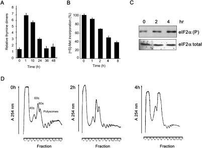
Figure 1.
Inhibition of protein synthesis following UVB-induced DNA damage. HeLa cells were mock- or UVB-irradiated (275 J/m2) and harvested at the times shown following exposure. (A) The relative amount of cyclo-pyrimidine dimers (CPDs) produced by this treatment were measured by ELISA using a monoclonal antibody specific to CPDs. Measurements are the mean of four independent experiments, and error bars represent 1 standard deviation from the mean. (B) Protein synthesis rates were determined by liquid scintillation counting of newly incorporated [35S]-methionine at the time points indicated. Measurements are the mean of three independent experiments normalized to that of the unirradiated cells. Error bars represent 1 standard deviation from the mean. (C) Cell lysates derived from control and UVB-irradiated cells were separated by SDS-PAGE, immunoblotted, and probed with antibodies against eIF2α. There was a significant change in the phosphorylation status of eIF2α but no other eIFs (see Supplemental Fig. S1C). (D) Cell lysates were separated on (10%–50%) sucrose density gradients and the absorbance across the gradient read at 254 nm. Positions of the 40S, 60S ribosomal subunits and polysomal fractions are indicated.

