Figure 2.
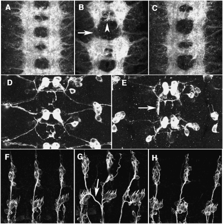
MID is required for CNS and PNS pathfinding. (A–C) A stage 16 embryo stained with the monoclonal antibody (mAb) BP102 to reveal the axonal scaffold of the CNS. Anterior is up. (A) Normal CNS. (B) In mid1 mutant, the longitudinal tracts were interrupted (arrow) and posterior commissures were thinner (arrowhead). (C) Normal axonal scaffold is restored by pan-neural expression of MID (elav-Gal4/+; mid1/mid1; UAS-mid/+ embryo). (D,E) Visualization of lim3A+ motor neurons in a stage16 embryo using the lim3A-tau-myc transgene (anti-Myc epitope staining). (D) Normal. (E) Motor axons stall and follow a longitudinal path in mid1 mutant (arrow). Similar motoneuron projection defects were observed in mid2 and midD62 embryos (data not shown). (F–H) A stage16 embryo stained with mAb 22C10 to reveal the sensory axons in the PNS. Anterior is left and dorsal up. (F) Normal PNS. (G) In mid1 mutant, sensory axons branch excessively or cross the segment borders (arrow). (H) Restored dorsal sensory axons in an elav-Gal4/+; mid1/mid1; UAS-mid/+ embryo. Similar phenotypes of CNS and PNS were also observed in mid2 or midD62 mutant embryos (data not shown).

