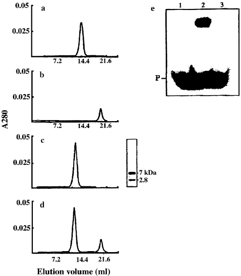
Figure 5.
Interaction of Sso7d with melittin, and DNA-binding activity of the melittin–Sso7d complex. Superdex-75 gel filtration columns loaded with 0.5 ml of 10 mM Tris-HCl pH 7.5 containing: (a) 35 µM Sso7d; (b) 35 µM melittin; (c) 35 µM Sso7d and 35 µM melittin; or (d) 35 µM Sso7d and 70 µM melittin after incubation at 50 °C for 30 min. Columns were eluted with 10 mM Tris-HCl pH 7.5, 0.2 M NaCl at a flow rate of 18 ml h–1. The SDS-PAGE analysis of the peak from column (c) (5 µg) is reported; after electrophoresis, the gel was silver-stained. (e) Sso7d (Lane 2) and the melittin–Sso7d complex (Lane 3) (1.5 µg of protein per lane) were utilized for the band-shift assay performed as described in Materials and methods; Lane 1 contains the double-stranded DNA probe only (P). The melittin–Sso7d complex was prepared as described in Materials and methods.

