Figure 2.
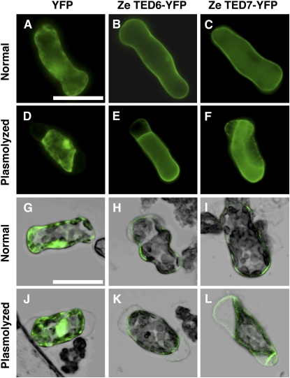
Localization of TED6- and TED7-YFP Fusion Proteins in Zinnia Mesophyll Cells.
Normal fluorescent images ([A] to [F]) and laser scanning confocal images merged with bright-field images ([G] to [L]) of cells cultured for 48 h after electroporation. Cells were observed before ([A] to [C] and [G] to [I]) or after plasmolysis ([D] to [F] and [J] to [L]) in medium supplemented with twofold mannitol content of the normal medium. Bars in (A) for (A) to (F) and in (G) for (G) to (L) = 50 μm.
(A), (D), (G), and (J) Cells electroporated with 35SPro:YFP.
(B), (E), (H), and (K) Cells electroporated with 35SPro:Ze TED6-YFP.
(C), (F), (I), and (L) Cells electroporated with 35SPro:Ze TED7-YFP.

