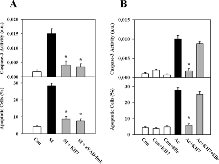
FIGURE 1.
Apoptosis of EC is abolished by suppression of sAC. A, exposure to simulated ischemia (SI; glucose-free anoxia at pH 6.4) induced an increase in caspase-3 activity and the number of apoptotic cells (Hoechst 33342 staining of nuclei) compared with control cells (Con), which was suppressed by inhibition of sAC (10 μmol/liter KH7). A similar effect was found upon treatment with the pan-caspase inhibitor benzyloxycarbonyl-VAD-fluoromethyl ketone (zVAD-fmk; 50 μmol/liter). B, exposure to acidosis (Ac) induced an increase in caspase-3 activity and the number of apoptotic cells (Hoechst 33342 staining of nuclei), which was suppressed by inhibition of sAC (10 μmol/liter KH7). The effects of KH7 treatment were abolished by addition of the cAMP analog 8-bromo-cAMP (8Br; 30 μmol/liter). Values are means ± S.E. (n = 7–11). *, p < 0.05 versus simulated ischemia or acidosis. a.u., arbitrary units.

