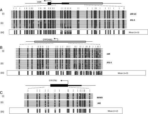
FIGURE 6.
Methylation analysis of vitamin D-associated genes in CCA-derived trophoblast cell lines. High levels of methylation were detected for both VDR (A) and CYP27B1 (C) in choriocarcinoma cell lines (n = 2). All choriocarcinoma cell lines (n = 3; BeWo not shown) examined showed hypermethylation of the CYP24A1 gene relative to full term placental tissue or purified first trimester trophoblasts. Numbers of each type of tissue are listed in parentheses. Between 8 and 12 individual clones were sequenced for each sample. Circles, CpG sites within the assayed region. Closed circles, methylation; open circles, lack of methylation. Missing circles indicate CpG sites for which no information was obtained. Gray boxes correspond to GpG island locations, and arrows denote start site of translation within exon 1 (black line). iii, mean methylation levels seen at each CpG site for each type of sample tested.

