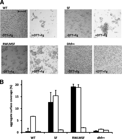
FIGURE 3.
Fibrinogen-induced CHO cell aggregation. CHO cells expressing WT and mutant αIIbβ3 receptors (CHO-αIIb/β3(SF) (SF) and CHO-αIIb(RWLM)/β3(SF) (RWLMSF)) and dhfr+ cells were washed with Tyrode's buffer and incubated in the presence or absence of DTT for 20 min at room temperature. Cells were resuspended at 3.75 × 10⁶ cells/ml in Tyrode's buffer containing 1 mmol/liter CaCl2 in the presence or absence of 0.5 mg/ml fibrinogen and were rotated for 20 min. Cells were fixed before analysis. A, the images are representative of four individual experiments performed in the presence of both DTT and fibrinogen (+DTT+Fg) or in the absence of DTT and presence of fibrinogen (-DTT+Fg). Scale bar = 100 μm. B, shown are the results from quantitative analysis of CHO cell aggregation presented in A in the absence of DTT and presence of fibrinogen (black bars), in the presence of both DTT and fibrinogen (white bars), and in the absence of both DTT and fibrinogen (gray bars). The aggregate surface coverage corresponds to the area covered by all aggregates in one view field (1807 × 1419 μm) normalized to the total surface measured. Data are the means ± S.E. of four individual experiments. In each experiment, four view fields were analyzed for each condition.

