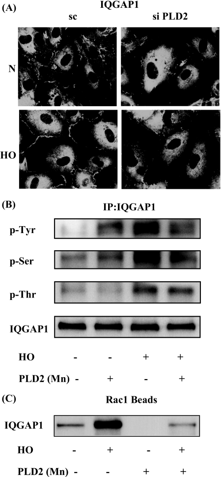
FIGURE 8.
PLD2 regulates hyperoxia-induced IQGAP1 activation and association with Rac1. A, HPAECs were transfected with scrambled or PLD2 siRNA, exposed to either normoxia or hyperoxia (3 h), and probed with anti-IQGAP1 antibody. Redistribution of IQGAP1 was examined by immunofluorescence microscopy using a ×60 oil objective. Shown is a representative immunofluorescence micrograph from three independent experiments. B, HPAECs were infected with vector control or adenoviral construct of mPLD2 mutant (Mn), exposed to either normoxia or hyperoxia (3 h), cell lysates were subjected to immunoprecipitation with anti-IQGAP1 antibody, and immunoprecipitates were assayed for phosphorylation of IQGAP1 with anti-phosphoserine, anti-phosphothreonine, or anti-phosphotyrosine antibodies as described under “Experimental Procedures.” Shown is a representative Western blot from three independent experiments. C, cell lysates from B were subjected to immunoprecipitation with PAK-1 PBD-agarose beads to bring down activated Rac1 bound to GTP as described under “Experimental Procedures.” Rac1-GTP bound to PAK-1 PBD was separated by 4-20% SDS-PAGE and probed with anti-Rac1 antibody. Shown is a representative blot from three independent experiments.

