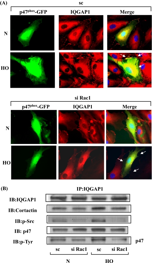
FIGURE 9.
Rac1 siRNA attenuates hyperoxia-induced IQGAP1 redistribution to cell periphery and co-localization with p47phox. A, HPAECs were transfected with scrambled or Rac1 siRNA, and additionally transfected with plasmid DNA of p47phox-GFP. 24 h later cells were exposed to either normoxia or hyperoxia (3 h) and analyzed by immunofluorescence microscopy for IQGAP1 (red) and p47phox-GFP (green) localization. In normoxic condition, IQGAP1 and p47phox-GFP were distributed diffusely in the cytosol and hyperoxia induced redistribution of both the proteins to cell periphery, where they appear to co-localize (yellow in merged image). Rac1 siRNA blocked the redistribution and co-localization of IQGAP1 and p47phox (less yellow visible in merged image). A representative image from three independent experiments is shown. B, in parallel experiments, HPAECs were transfected with scrambled or Rac1 siRNA, cells exposed to either normoxia or hyperoxia (3 h), and total cell lysates were subjected to immunoprecipitation with anti-IQGAP1 antibody as described under “Experimental Procedures.” Immunoprecipitates were analyzed after separation by SDS-PAGE for total IQGAP1, cortactin, Src, and p47phox with anti-IQGAP1, -cortactin, -Src, and -p47phox antibodies, respectively. Shown is a representative blot from three independent experiments.

