Fig. 1.
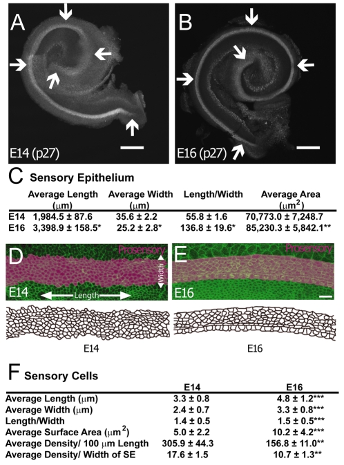
Convergence and extension of the cochlear prosensory domain. (A,B) Whole mounts of the cochlea at E14 (A) and E16 (B). The prosensory domain is marked by expression of p27Kip1 (arrows). Note the extension and narrowing of the prosensory domain (arrows) that occurs between E14 and E16. (C) Comparison of changes in length, width, length/width ratio and surface area of the sensory epithelium between E14 and E16. There is a significant increase in length, a significant decrease in width and a significant increase in overall area. (D,E) Phalloidin labeling (green) of cell-cell boundaries in the cochlear duct. The prosensory domain is illustrated in violet. Arrows in D indicate orientation of length and width axes. The lower aspect of each panel illustrates outlines for individual cells within the prosensory domain. (F) Comparison of changes in length, width, length/width ratio, surface area, and cell density per unit length and across the width of the sensory epithelium between E14 and E16. The decrease in density of cells along the width of the epithelium is consistent with ongoing convergence. Scale bars: 200 μm in A,B; 10 μm in E (same magnification in D). *P<0.005, **P<0.05, ***P<0.0001.

