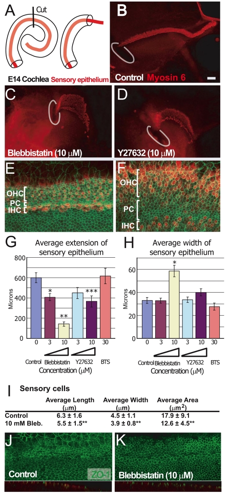
Fig. 5.
Inhibition of myosin II activity disrupts cochlear CE. (A) Outgrowth of the sensory epithelium was assayed by cutting the intact cochlear duct at its midpoint and then maintaining the basal piece in vitro. Over the subsequent 72-96 hours the sensory epithelium extended outwards from the cut end of the explant. (B) Control explant established as described in A. The site of the initial cut is marked in white. The sensory epithelium (hair cells marked in red) extends from the initial cut site. (C) Explant established as in B, but treated with 10 μM of the myosin II inhibitor blebbistatin beginning at E14. The sensory epithelium is noticeably shorter and wider. (D) An explant established as in B, but treated with 10 μM of the ROCK inhibitor Y27632. The phenotype of the sensory epithelium is similar to that in C. (E,F) Images of the sensory epithelium from control (E) and 10 μM blebbistatin-treated (F) explants labeled with anti-myosin 6 (red) and phalloidin (green). Additional inner pillar cells (PC) are present in blebbistatin-treated explants. Patterning in the IHCs and OHCs is also disrupted. Abbreviations as in Fig. 2. (G) Treatment with either blebbistatin or Y27632 results in a significant and dose-dependent decrease in extension of the sensory epithelium. By contrast, the skeletal muscle myosin inhibitor BTS did not affect extension. (H) Treatment with 10 μM blebbistatin leads to a significant increase in the width of the sensory epithelium. However, the width of the sensory epithelium was not significantly increased in explants treated with 10 μM Y27632. (I) Blebbistatin treatment leads to changes in cell shape that are consistent with changes in Myh10DN mutants. (J,K) Surface (upper panel) and cross-section (lower panel) views of control (J) and blebbistatin-treated (K) explants labeled with an antibody specific for the tight junction protein ZO1 (green). Tight junctions are present in explants from both conditions and are restricted to the lumenal surfaces. Scale bars: 50 μm in B (same magnification in C,D); 20 μm in E (same magnification in F,I,J). *P<0.005, **P<0.001, ***P<0.01.

