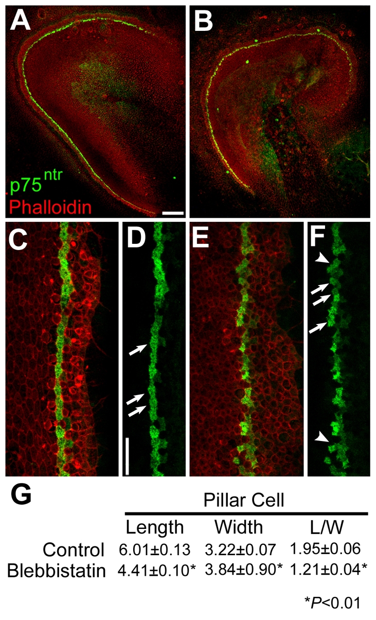
Fig. 6.

Pillar cell shape is affected in blebbistatin-treated cochlear explants. (A) Low magnification view of a control explant established at E13 and fixed after 72 hours in vitro. The pillar cells (green) extend in a single line along the length of the explant. (B) Low magnification view of an explant established and labeled as in A, but treated with 10 μM blebbistatin for 48 hours beginning after 24 hours in vitro. The overall length of the epithelium is shorter and the row of pillar cells appears less organized. (C) High magnification view of the sensory epithelium from a control explant labeled as in A. The pillar cells are present in a single organized row. (D) The same view as in C with the red channel removed. Pillar cells are indicated (arrows). (E) High magnification view of the sensory epithelium from an explant treated with 10 μM blebbistatin, labeled as in A. (F) The same view as in E, with the red channel removed. Note the poor organization of the row of pillar cells and the more rounded shape. (G) Quantification of the average length and width of pillar cells in control and blebbistatin-treated explants. In blebbistatin-treated explants, pillar cell lengths are significantly shorter, whereas pillar cell widths are significantly greater, resulting in a significant decrease in the length/width ratio. Scale bars: 100 μm in A (same magnification in B); 20 μm in D (same magnification in C,E,F).
