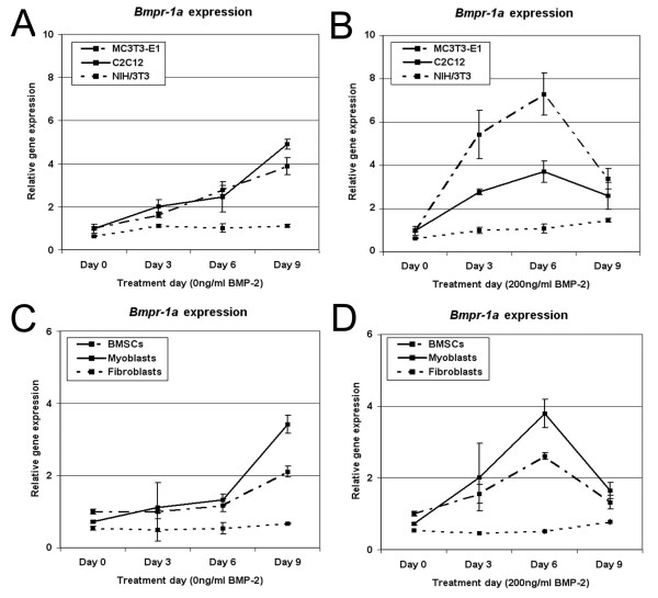
Figure 4.
Cellular response to BMP-2 correlates to theexpression levels of Bmpr-1a. Bmpr-1a expression levels were examined using qPCR and normalized to the housekeeping gene Gapdh. Values were generated for cell lines grown in the absence of BMP-2 (A) or with BMP-2 added (B), and in mouse primary derived cells grown without (C) or with (D) BMP-2 treatment. Bmpr-1a expression increased over time when cells were grown in osteogenic media, and were even greater under BMP-2 stimulation. The lowest Bmpr-1a levels were observed in NIH/3T3 cells and primary fibroblasts, which were previously shown to be the least sensitive to BMP-2 treatment.

