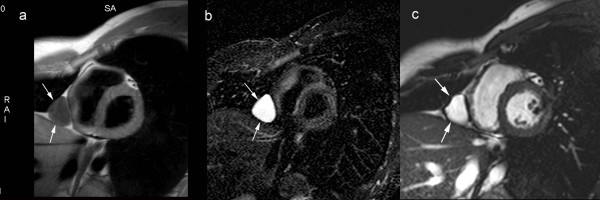
Figure 2.
Congenital pericardial cyst. T1-weighted spin-echo CMR. (a) T2-weighted short-tau inversion-recovery spin-echo CMR (b), and cine CMR (c) in cardiac short-axis. The pericardial cyst is visible as a well-delineated, homogeneous soft-tissue structure (arrows) along the right paracardiac border implanted on a normal pericardium. The cyst content typically has signal characteristics of water on CMR.

