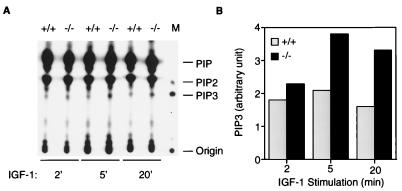
Figure 3.
PIP3 accumulation in Pten+/+ and Pten−/− cells. (A) PIP3 levels in Pten+/+ and Pten−/− ES cells after IGF-I stimulation. Cells were starved in a serum-free medium for 16 hr, and then labeled with [32P]orthophosphate (0.5 mCi/ml) for 2 hr. Cells then were stimulated by IGF-I (1 μg/ml) for 2, 5, or 20 min before harvesting. Phospholipids were extracted and analyzed on a TLC plate. Assignment of PIP, PIP2, and PIP3 was done according to in vitro 32P-labeled phosphoinositides standards (see Materials and Methods). In lane M, [32P]-labeled PIP3 is shown as a marker. (B) Quantitation of PIP3 levels in Pten+/+ and Pten−/− ES cells after IGF-I stimulation. The amount of radioactivity corresponding to PIP3 was measured with a PhosphorImager and presented as an arbitrary unit.

