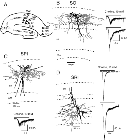
Fig. 1.
Type IA currents in various interneurons in the CA1 region of guinea pig hippocampal slices. A, scheme of a hippocampal slice illustrating the approximate location of various interneuron types studied in the CA1 region. B, Neurolucida drawing of a biocytin-filled SOI showing that the axon is primarily projecting in the SO region. Sample recording of type IA current recorded from two SOIs are shown on the right. C, Neurolucida drawing of a biocytin-filled SPI showing that the axon is primarily projecting in the pyramidal cell layer. Sample recording of type IA current recorded from SPIs is shown at the bottom. D, Neurolucida drawing of a biocytin-filled SRI showing that the axon is targeted primarily to the SR region. Sample recording of type IA currents recorded from SRIs either decayed completely during agonist pulse (top trace) or had some steady-state current at the end of the agonist pulse (bottom trace). Dendrites are shown in black and axons in gray in all Neurolucida drawings.

