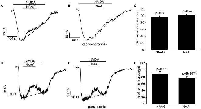
Figure 4.
Test of block of NMDA-evoked current at –74 mV by NAAG and NAA in cerebellar white matter oligodendrocytes (A–C) and granule cells (D–F). (A) Representative trace shows block of 60 µM NMDA-evoked response by 1 mM NAAG in oligodendrocyte. (B) Representative trace shows block of 60 µM NMDA-evoked response by 1 mM NAA in oligodendrocyte. (C) Mean effect of 1 mM NAAG (five cells) and 1 mM NAA (three cells) on the NMDA-evoked current, calculated assuming for simplicity that desensitization of the NMDA-evoked current follows a linear time course (dashed lines in A and B: the NMDA-evoked current at the end of the NAAG or NAA application was divided by the current predicted assuming linear desensitization). (D) Cell showing block of 60 µM NMDA-evoked response by 1 mM NAAG in granule cell (not all cells showed any block). (E) Cell showing block of 60 µM NMDA-evoked response by 1 mM NAA in granule cell (not all cells showed any block). (F) Mean effect of 1 mM NAAG (nine cells) and 1 mM NAA (five cells) on the NMDA-evoked current, calculated assuming for simplicity that desensitization of the NMDA-evoked current follows a linear time course.

