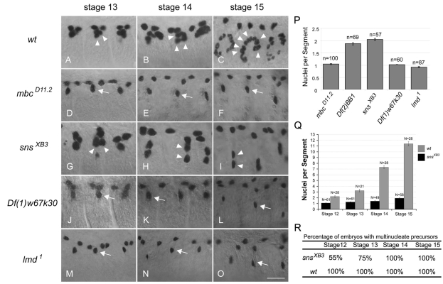
Fig. 1.
Fusion of the Eve-expressing DA1 founder cell in various mutant embryos. (A-O) Abdominal segments 3-5 are shown in embryos stained with anti-Eve to mark the nuclei of the DA1 muscle and two pericardial cells per hemisegment. (A-C) Wild-type, (D-F) mbcD11.2/mbcD11.2, (G-I) snsXB3/snsXB3, (J-L) Df(1)w67k30/Y and (M-O) lmd1/lmd1. The founder cell for DA1 remains mononucleate in embryos lacking mbc, kirre and rst [Df(1)w67k30], or lmd at developmental stages when significant fusion is observed in wild-type embryos (arrows). In embryos mutant for sns, by contrast, the Eve-expressing DA1 founder cell undergoes limited fusion to generate bi- or tri-nucleate syncitia (arrowheads). (P) The average number of DA1 nuclei per hemisegment was quantitated in late stage 15 embryos of each mutant genotype. (Q) The fusion profile of precursor formation in wild-type and snsXB3 embryos shown as the average number of DA1 nuclei per hemisegment. (R) The percentage of embryos observed with any hemisegments showing DA1 precursor formation. Scale bar: 20 μm.

