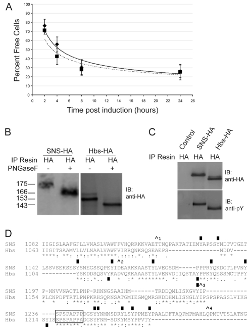
Fig. 3.
Hbs and Sns have comparable aggregation behavior with Kirre-expressing cells and undergo similar biochemical modifications. (A) Kinetics of trans-heterotypic aggregation of S2 cells expressing Kirre co-cultured with S2 cells expressing Sns (diamonds, bold line) and S2 cells expressing Hbs (squares, dashed line). (B,C) Sns-HA or Hbs-HA expressed pan-mesodermally with mef2Gal4 and immunoprecipitated with anti-HA resin. (B) Immunoblots of untreated (Mock) or PNGaseF-treated Sns-HA and Hbs-HA reveals modification by N-linked glycans. (C) Immunoblots of Sns-HA, Hbs-HA or control (mef2Gal4/+) samples probed with anti-HA or anti-phosphotyrosine antibodies. (D) Alignment of transmembrane and cytodomain sequences from Sns and Hbs. The entire Hbs cytodomain is shown. Only the membrane proximal half of the Sns cytodomain is shown. The caret positions correspond to (1) AA1113, (2) AA1164, (3) AA1232 and (4) AA1278 of SNS, and served as deletion endpoints in a structure/function analysis of Sns. Conserved sequences from Hbs, important PxxP motifs and tyrosine residues crucial for Sns function are highlighted.

