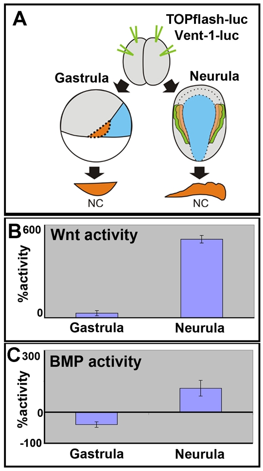
Fig. 7.

Changes in Wnt and BMP activity during NC development. (A) Two-cell stage embryo received animal and equatorial injections of the indicated reporter constructs. The NC was dissected at the gastrula or neurula stages and luciferase was measured. (B) Measurements of canonical Wnt activity using the TOPflash luciferase reporter in NC explants. Activity expressed as the percentage of activity seen in stage 10 NC explants. (C) Measurements of BMP activity using the Vent1 luciferase reporter in NC explants. Activity was determined as percentage of activity in epidermal explants of the same stage and shown normalised to activity in the epidermis.
