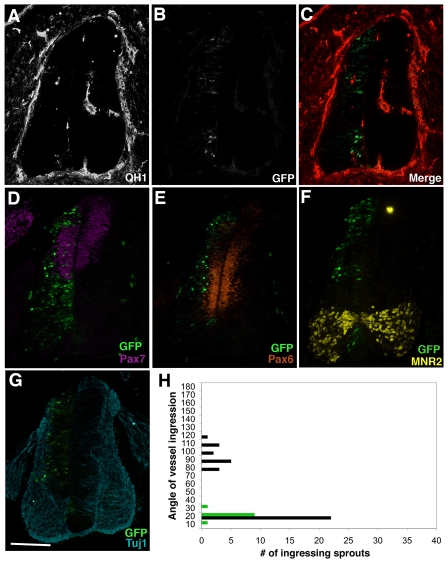
Fig. 6.
VEGF signaling from the neural tube is required for blood vessel ingression. Quail neural tubes were electroporated with sFlt1-GFP and analyzed as previously described. (A-C) No medial vessel ingression and little ventral vessel ingression was seen in areas of the neural tube that were eGFP positive. (D-G) Neural patterning is not detectably perturbed on the electroporated side of the neural tube (left) based on Pax7 (D, purple), Pax6 (E, orange), MNR2 (F, yellow) and Tuj1 (G, blue) expression patterns. (H) Quantitative analysis of five electroporated neural tubes showed no medial and few ventral vessel ingression points in areas of localized sFlt1 expression (green), compared with the control contralateral side (black) (n=5 embryos). Scale bar: 100 μm.

