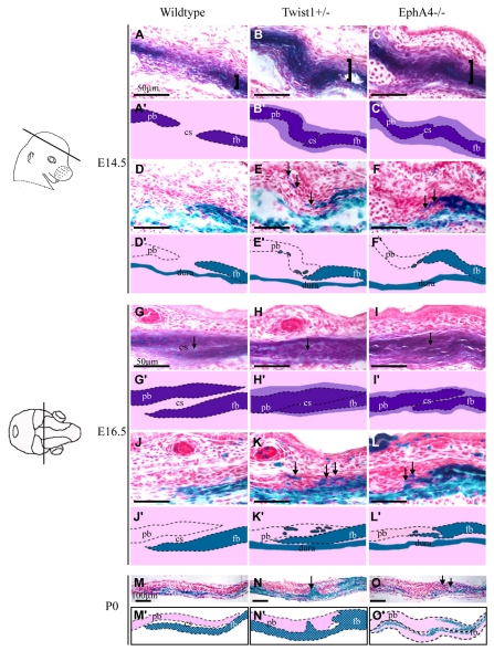
Fig. 2.
Increased number of alkaline phosphatase-expressing cells and defect in the neural crest-mesoderm boundary in coronal sutures of EphA4-/- embryos. Heads of wild-type Wnt1-Cre; R26R, Twist1+/-; Wnt1-Cre; R26R, and EphA4-/-; Wnt1-Cre; R26R embryos at E14.5, E16.5 were sectioned in the plane indicated and alternate sections were stained either for alkaline phosphatase (A-C and G-I) or lacZ expression (D-F and J-L). Pups at P0 were examined only for lacZ expression (M-O). Schematics depicting key results are shown below each image (A′-O′). Note widening of ALP domain (A-C, brackets) and expansion of ALP expression into suture (G-I, arrows). Also note lacZ-positive (neural crest) cells located ectopically in prospective coronal suture and parietal bone (D-F,J-L,M-O, arrows). cs, coronal suture; fb, frontal bone; pb, parietal bone.

