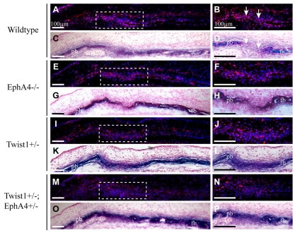
Fig. 6.
Altered distribution of P-Smad1/5/8-expressing cells in coronal sutures of Twist1 and EphA4 individual and combination mutant embryos. Heads of E14.5 embryos were sectioned as in Fig. 2 and stained for P-Smad1/5/8 activity. Alternate sections were stained for ALP. Right panels show enlargements of dashed squares in left panels. Note concentration of stained nuclei in osteogenic fronts of wild-type embryos (A-D). In mutant embryos, note scattered stained nuclei and loss of concentration of stained nuclei in osteogenic fronts (E-P). fb, frontal bone; pb, parietal bone.

