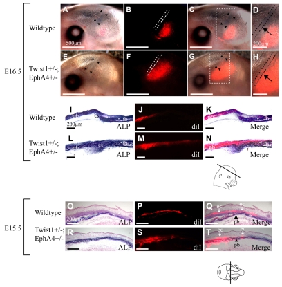
Fig. 8.
Mis-migration of osteogenic precursor cells in Twist1+/-; EphA4+/- mutant embryos. We injected DiI into parietal bone rudiments of E13.5 wild-type (A-D,I-K,O-Q) and mutant (E-H,L-N,R-T) embryos, in vivo. Embryos were allowed to develop exo utero to E16.5 (A-N) or E15.5 (O-T), when they were examined for the distribution of DiI by epifluorescence microscopy. Sections shown in I,K,L,N,O,Q,R and T were also stained for ALP activity. E16.5 (A-N): embryos shown in whole mount in A-H were sectioned; images of the sections are shown in I-N. (A,E) Brightfield images of whole mounts. Note location of coronal sutures (arrowheads). (B,F) Epifluorescence images, with dotted lines showing location of sutures. (C,G) Merged brightfield and epifluorescence images. Area within dashed square is magnified in D,H. Labeled osteogenic precursor cells migrate apically (B-D) and insert into the growing bone (J,K). Note DiI-labeled cells are excluded from coronal sutures of wild-type (B,D, dotted lines) but not mutant embryos (G,H arrows). This is also clearly evident in sections: compare J,K with M,N. E15.5 (O-T): images are from coronal sections of E15.5 embryos (plane of section indicated). (O,R) Brightfield images stained for ALP; (P,S) epifluorescence images; (Q,T) combined brightfield and epifluorescence images. Note that in wild-type embryos, labeled cells are located largely in layer flanking (ectocranial to) the osteogenic layer (arrows, `ec'). In mutant embryos, labeled cells are located diffusely in and around osteogenic layer of parietal bone (arrowheads, `pb'). Closely similar results were obtained in more than ten injected wild-type embryos and three injected mutant embryos.

