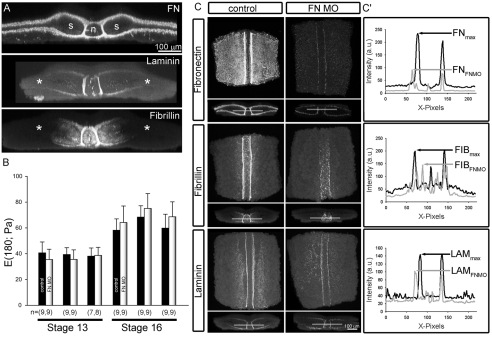
Fig. 5.
Role of ECM in tissue stiffness. (A) Representative transverse maximum projections of confocal sections show fibronectin (FN), laminin (LAM), and fibrillin (FIB) are all present within the dorsal tissues by stage 16. FN surrounds both axial (n, notochord) and paraxial mesoderm (s, prospective somitic mesoderm). LAM and FIB surround the axial mesoderm but do not assemble more laterally (asterisk). (B) The stiffness of explants injected with fibronectin morpholinos (FNMO) does not differ from uninjected explants at either stage 13 or at stage 16. Seven to nine explants from each of three separate clutches of embryos were tested. (C) Representative maximum projections of FNMO-injected dorsal isolates exhibit severe reduction in fibronectin fibrils and also show defects in assembly of fibrillin and laminin compared with control isolates sectioned with identical confocal settings. (C′) Intensity profiles collected mediolaterally across the midline (indicated by transparent line across the midline in xz-projections shown in C indicate as expected that FN assembly is severely reduced but FIB and LAM also exhibit significant reductions.

