
Fig. 1.
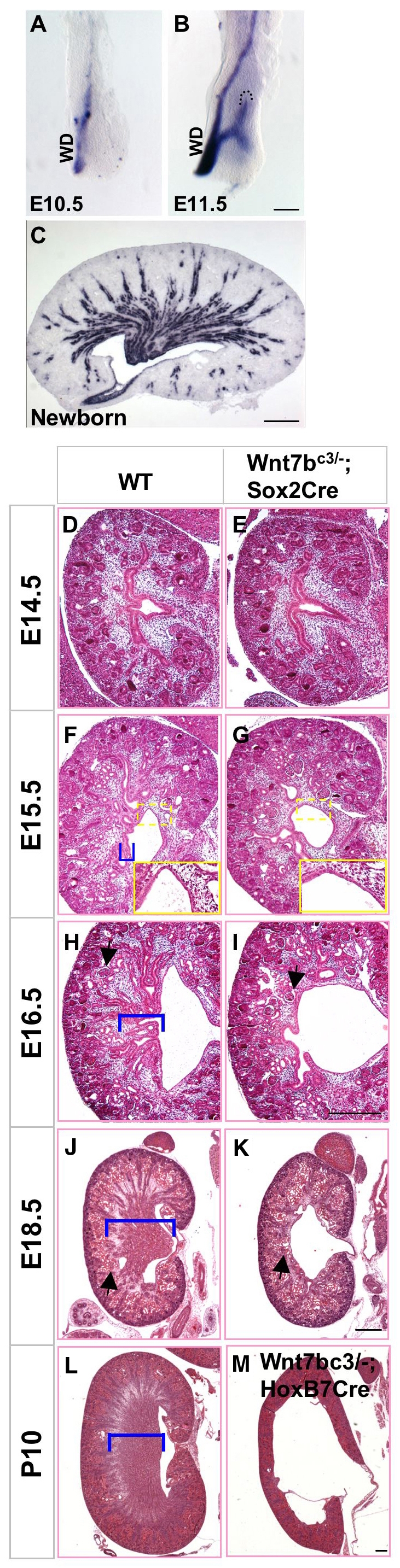
Wnt7b is essential for the development of the medullary component of the mouse kidney. (A-C) In situ hybridization analysis of Wnt7b expression in the developing mouse kidney. Prior to ureteric bud outgrowth, Wnt7b mRNA was expressed at low levels in the mesonephric/Wolffian duct (WD) epithelium (A). After ureteric bud invasion into the metanephric mesenchyme, Wnt7b expression was restricted to the ureteric trunk epithelium (B) and its derivatives, the collecting duct epithelium and ureter of the kidney (C). Expression was not observed at the ureteric tips (boundary demarcated by dashed lines in B). Scale bars: 100 μm in A,B; 400 μm in C. (D-M) Hematoxylin and Eosin staining of kidney sections from wild-type littermates (D,F,H,J,L) and Wnt7b mutants (E,G,I,K,M) as indicated. The renal medullary compartment (bracketed) was first evident at E15.5 in wild-type embryos, but was absent from mutants at this and all later stages. The renal pelvis appeared normal (F and G, insets). Arrows in I and K point to renal corpuscles adjacent to the pelvic space. Arrows in panels H and J point to renal corpuscles that lie above the renal medulla. The P10 ureteric epithelium-specific Wnt7b mutants (M) exhibited hydroureter and hydronephrosis. Scale bars: 200 μm.