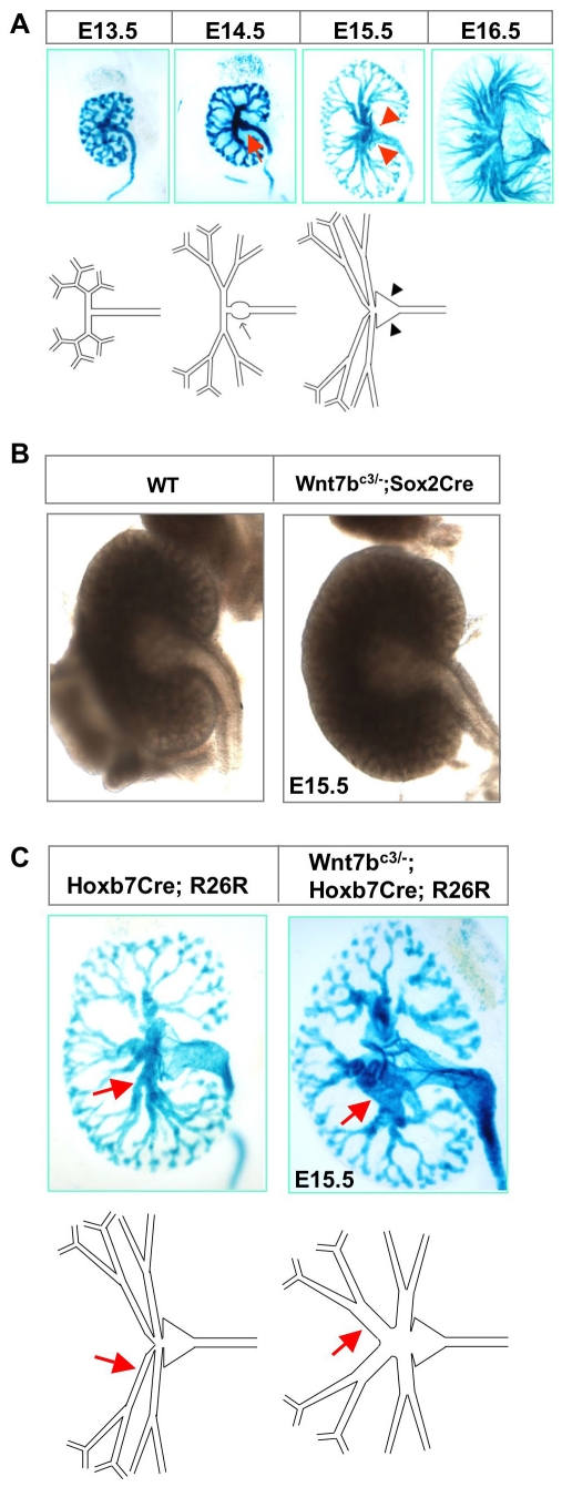
Fig. 3.
Morphogenesis of the distal collecting duct epithelium is disrupted in Wnt7b mutants. (A) The organization of the developing collecting duct network was visualized through ureteric epithelium-specific, histochemical staining for E. coli β-galactosidase activity in wild-type kidneys from Hoxb7Cre;R26R embryos. Data represent thick (150-300 μm) vibratome sections at the stages indicated and are schematized in the panel below. Arrows highlight a swelling at the intersection between the ureter and the collecting duct epithelium; arrowheads indicate the triangular-shaped renal pelvis. (B) An analysis of freshly dissected kidneys showed that the ureter was not dilated in Wnt7b mutants at E15.5. (C) By contrast, the collecting duct epithelium was dilated in the prospective medullary region of Wnt7b mutants at E15.5 when the R26R reporter was activated in the context of the collecting duct network of the Wnt7b mutant kidney. Arrows indicate prospective medullary collecting ducts.

