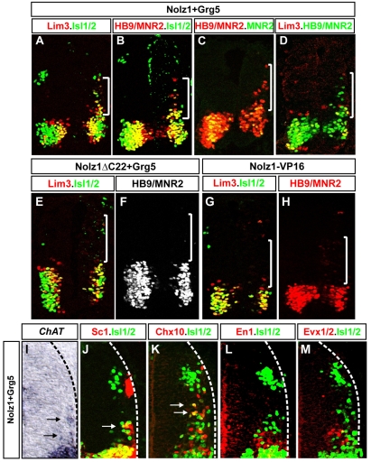
Fig. 6.
Nolz1-Grg5 complexes induce postmitotic MNs. Transverse sections of St 21 embryonic chick spinal cords electroporated on the right. White brackets denote ectopic MNs. (A-H,J-M) Immunohistochemical analyses using antibodies against MN and ventral interneuron markers. Arrows indicate ectopic MNs expressing the MN surface marker SC1 (J) or Chx10 (K). (I) Arrows mark ectopic MNs expressing transcripts for choline acetyltransferase (ChAT). Dashed lines mark the lateral extent of the spinal cord.

