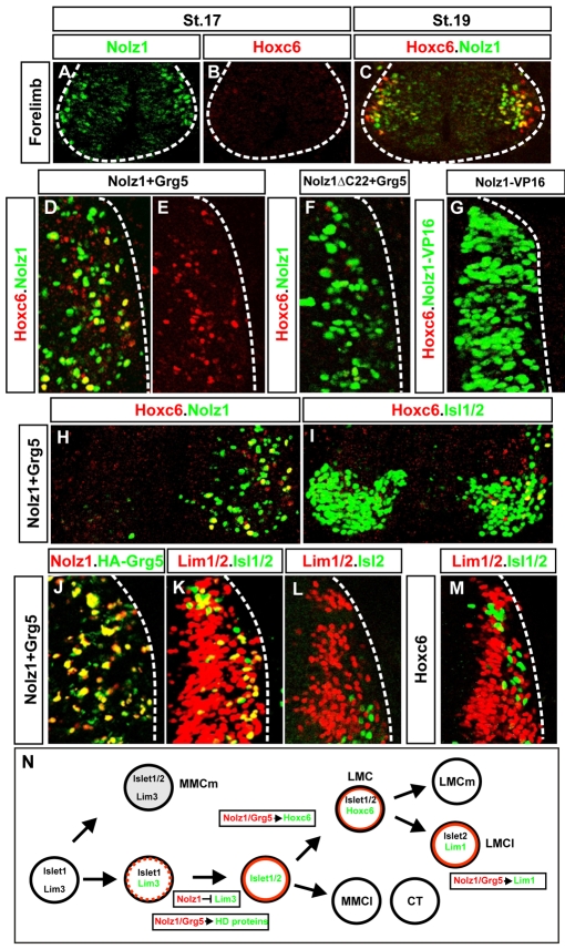
Fig. 8.
Coexpression of Nolz1 and Grg5 induces Hoxc6 expression and partial lateral LMC specification. (A-C) Confocal micrographs of ventral chick embryonic spinal cords showing that the onset of Nolz1 expression occurs prior to that of Hoxc6. The dashed line outlines the spinal cord. (D-G) Confocal micrographs of electroporated dorsal right halves of St 23 chick spinal cords. Grg5 expression is not shown but is ∼95-98% coincident with exogenous Nolz1. The dashed line marks the lateral boundary of the spinal cord. (H,I) Confocal micrographs of the ventral MN domain of St 23/24 chick embryonic spinal cords electroporated on the right. Yellow cells in panel I mark thoracic MNs that express Hoxc6. (J-M) Confocal micrographs of electroporated dorsal right halves of St 23 chick spinal cords. The dashed line marks the lateral boundary of the spinal cord. The right-hand side is electroporated in all cases. The high levels of Nolz1 elicited by overexpression preclude the detection of endogenous Nolz1 under these imaging conditions. (N) Model for Nolz1-dependent specification of MN subtype identity. Orange lines mark cells expressing Nolz1. Broken orange line refers to transient Nolz1 expression. Nolz1 downregulates Lim3 expression in prospective Lim3-negative MNs and, in a complex with Grg5, maintains the expression of key MN determinants. At limb levels, Nolz1-Grg5 complexes confer forelimb LMC identity through induction of Hoxc6, and induce Lim1 expression in prospective LMCl MNs.

