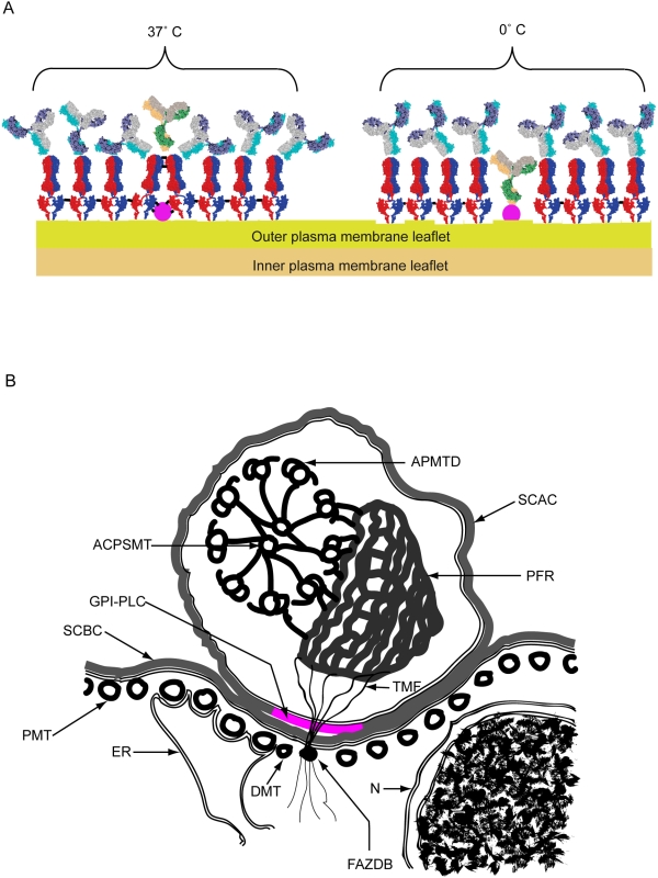
Figure 8. Scheme 1. Cartoons showing the arrangement of VSG dimers shielding the GPI-PLC at 37°C and allowing access to the GPI-PLC at 0°C as well as the cross-sectional relationships between the GPI-PLC and other surrounding structures.
Panel A. Cartoon of the arrangement of VSG dimers (red & blue) on the outer leaflet of the plasma membrane of bloodstream form trypanosomes after mild cross-linking (black bars) of proteins by brief fixation (10 min) at either 37°C or 0°C. Subsequent access to the VSG is shown by the position of attached IgG anti-VSG (grey, mauve and turquoise) and access to a partially buried protein, such as the GPI-PLC (mauve), is shown by the position of attached or excluded IgG anti-GPI-PLC (yellow, green and grey). Panel B. Cartoon of a cross section through the cell body and flagellar compartment showing the relationships between the GPI-PLC and the flagellar attachment zone together with associated structures, including the ACPSMT, axonemal central pair single mictotubule; APMTD, axonemal peripheral microtubule doublet; DMT, diminished microtubule [29]; ER, loop of endoplasmic reticulum contacting the four constant microtubules within the flagellar attachment zone; FAZDB, flagellar attachment zone dense body; GPI-PLC (magenta), the glycosylphospatidyl-inositol phospholipase C; N, nucleus covered by inner and outer nuclear membranes; PMT, pellicular microtubule; PFR, paraflagellar rod; SCAC, surface coat covering the plasma membrane of the flagellar compartment; SCBC, surface coat covering the plasma membrane of the cell body compartment; TMF, one of the transmembrane fibrils connecting the paraflagellar rod to the flagellar attachment zone dense body.

