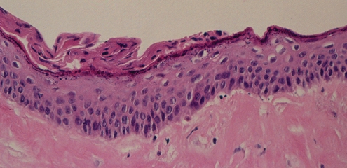
Figure 18).
Patient 5: Hematoxylin and eosin stain of the surface of the left capsule showing a thickened layer of squamous epithelium with early keratinization along the surface. This represents synovial metaplasia. Deep to this epithelial layer, there was a well-developed area of hyalinizing fibrosis that had a paucity of cells (original magnification ×250)

