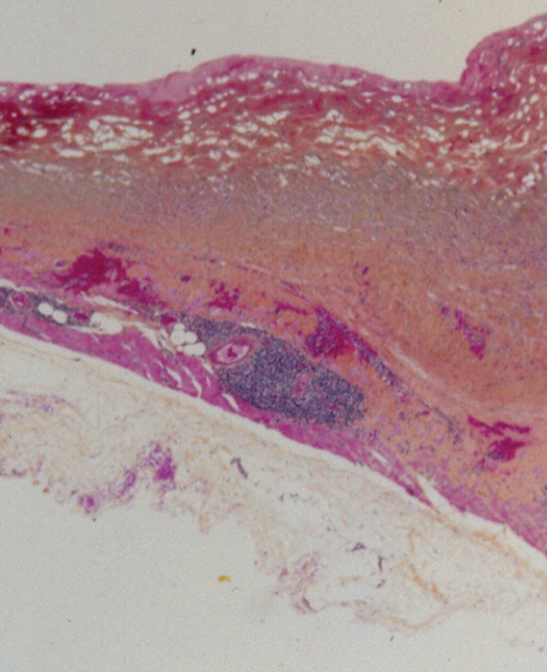
Figure 6).
Patient 2: World Health Organization stain showing a layer of organizing hematoma on the inner surface of the capsule. This was superimposed on organizing fibrovascular granulation tissue. The outer capsule was composed of collagen with random orientation. On the other aspect of the capsule, there were small aggregates of lymphocytes and evidence of recent bleeding. Note the degree of organization of this hematoma capsule. At four months, it was much more advanced than the hematoma in patient 1 at one month (Figure 2) (original magnification ×12.5)

